


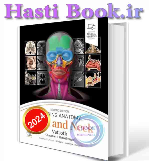
دانلود کتاب Imaging Anatomy: Head and Neck 2024 (ORIGINAL PDF)
نام کتاب:Imaging Anatomy: Head and Neck
نويسنده/نويسندگان: Surjith Vattoth MD FRCR
نوع فايل: pdf ORIGINAL
سال انتشار: 2024
تعداد صفحات : 664
ناشر :
Publisher :
شابک :9780443249648
نسخه چاپی تمام رنگی ، با کیفیت بالا ،جلد هارد
*بهترین کیفیت افست را از ما بخواهید*
شماره تماس ، واتس آپ و تلگرام 📞 09371686566📞
To buy in dollars, if you have PayPal, send a message to our Telegram
+989371686566
شماره تماس ، واتس آپ و تلگرام 📞 09371686566📞
🌸🌸🌸 🌸🌸🌸 🌸🌸🌸 🌸🌸🌸 🌸🌸🌸🌸
This richly illustrated and superbly organized text/atlas is an excellent point-of-care resource for practitioners at all levels of experience and training. Written by global leaders in the field, Imaging Anatomy: Head and Neck, second edition, provides a thorough understanding of the detailed normal anatomy that underlies contemporary imaging. This must-have reference employs a templated, highly formatted design; concise, bulleted text; and state-of-the-art images throughout that identify the clinical entities in each anatomic area, offering a unique opportunity to master the fundamentals of normal anatomy and accurately and efficiently recognize pathologic conditions.
- Features hundreds of detailed, full-color illustrations and more than 900 high-resolution, cross-sectional radiologic images that together illustrate the fine points of imaging anatomy for new and experienced head and neck imaging specialists
- Contains new chapters on external nose anatomy, the facial nerve in temporal bone, minor fissures and sutures around the temporal bone, and temporal bone anatomy on photon-counting detector (PCD) CT
- Provides updated, enlarged images and captions in areas such as facial muscles and the superficial musculoaponeurotic system, and frontal recess and related air cells
- Includes extensive new content on PCD CT; new details on relatively unknown anatomical foramina, such as the vomerovaginal canal and canaliculus innominatus; new content based on the International Frontal Sinus Anatomy Classification; and minute details on the course of nerves in the head and neck
- Includes a series of successive imaging slices in each standard plane of imaging (coronal, sagittal, and axial) to provide multiple views that further support learning
- Depicts common anatomic variants and covers the common pathological processes that manifest with alterations of normal anatomic landmarks
- Reflects new understandings of anatomy due to ongoing anatomic research as well as new, advanced imaging techniques
- Presents essential text in an easy-to-digest, bulleted format, enabling imaging specialists to find quick answers to anatomy questions encountered in daily practice
- Includes an eBook version that enables you to access all text, figures, and references with the ability to search, customize your content, make notes and highlights, and have content
.